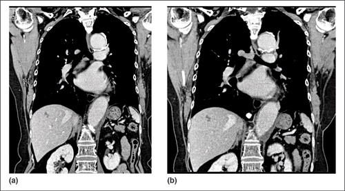
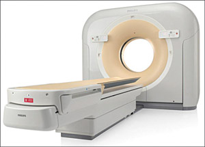
Diagnostic radiology tests utilising various imaging equipment like CT/X-ray are essential to arrive at an accurate diagnosis. iDose4 is an iterative-based reconstruction technology introduced by Philips in its CT scanners through which the scanner is enabled to reduce radiation dose up to 80 per cent without compromising on the quality of images. iDose4 is also capable of improving the image quality at low radiation dose. “This technology is amongst the most successful low-dose imaging technologies in the CT industry, which got widely appreciated by the entire radiology community,” says Raveendran Gandhi, senior director-Radiology, Philips Electronics, who shared more details on this technology.
Radiation—A concern and the reason behind iDose4
Talking about the effects of radiation exposure, Raveendran Gandhi said, “Exposure to ionising radiation leads to several biological side effects. Multiple clinical studies on radiation effects on foetus have consistently found an association between exposure to medical radiation during pregnancy and risk of childhood cancer in the offspring.”
According to Raveendran Gandhi, several clinical studies basing radiation effects on pediatric CT risks suggest that pediatric CT will result in significantly increased lifetime radiation risk over adult CT, both because of the increased dose per milliampere-second, and the increased lifetime risk per unit dose. In the UK, it has been estimated that approximately four per cent of diagnostic radiology procedures are CT examinations, but, their contribution to the collective radiation dose is approximately 40 per cent.
This emphasises the need for low-dose CT imaging technology.
iDose4—A different approach to image reconstruction
“For decades, filtered back projection (FBP) has been the industry standard for CT image reconstruction. While it is a very fast and fairly robust method, FBP is a sub-optimal algorithm choice for poorly sampled data or for cases where noise overwhelms the image signal. Such situations may occur in low-dose or tube-power-limited acquisitions, for instance in the scans of morbidly obese individuals,” says Raveendran Gandhi. Talking about the noise, he further adds, “Noise in CT projection data is dominated by photon count statistics. As the dose is lowered, the variance in the photon count statistics increases disproportionately. When this very high level of noise is propagated through the reconstruction algorithm, the result is an image with significant artefacts and high-quantum mottle noise.”
iDose4 is based on a completely different approach to image reconstruction (IR) techniques. Raveendran Gandhi says, “IR techniques attempt to formulate image reconstruction as an optimisation problem, i.e., IR attempts to find the image that is the ‘best fit’ to the acquired data. The noisiest measurements are given low weight in the iterative process; therefore they contribute very little to the final images.” He further adds, “Hence, IR techniques treat noise properly at very low signal levels, and consequently, reduce the noise and artefacts present in the reconstructed image. This results in an overall improvement of image quality at any given dose.” With IR techniques, the noise can be controlled for high spatial resolution reconstructions, hence, providing high-quality, low-contrast and spatial resolution within the same image. These are possible due to the recent innovations in hardware design and algorithm optimisations.
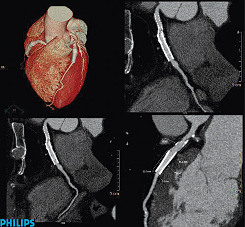
Working of iDose4. Whilst explaining the working of iDose4, a noteworthy point mentioned by Raveendran Gandhi is that in iDose4 algorithm, iterative processing is performed in both the projection and image domains. The reconstruction algorithm starts with projection data, where it identifies and corrects the noisiest CT measurements—those with very poor signal-to-noise (S/N) ratio, or very low photon counts. Each projection is examined for points that have likely resulted from very noisy measurements using a model that includes the true photons statistics. Through an iterative diffusion process, the noisy data is penalised and edges preserved. This process ensures that the gradients of underlying structures are retained, thus, preserving spatial resolution whilst allowing a significant noise reduction.
The noise that remains after this stage of algorithm is propagated to the image space. however, the propagated noise is now highly localised and can be effectively removed to support desired level of dose reduction. Then the iDose4 algorithm deals with subtraction of the image noise whilst preserving the underlying edges associated with true anatomy or pathology.


