
Unfortunately, fMRI, as it is used today, has a major drawback: It measures blood flow, or haemodynamics, which is an indirect measure of neural cell activity. It turns out that haemodynamics basically introduces a delay of five seconds. So fMRI is not able to detect fast variation.
Since neurons typically fire at an interval of the order of milliseconds, current fMRI techniques provide only a rough estimate of what the brain is doing at any given moment. fMRI scans also have a relatively low spatial resolution, measuring activity in areas of 100 microns—a volume that typically contains 10,000 neurons, each with varying activation patterns. Efforts to fine-tune fMRI have focused on developing stronger magnets.
Calcium tracking
Researchers have found that tracking calcium—a key messenger in the brain—may be a more precise way of measuring neural activity than current imaging techniques (viz, fMRI). When a neuron sends an electrical impulse to another neuron, calcium-specific channels in the neuron’s membrane instantaneously open up, letting calcium flow into the cell. It’s a very dramatic signal change.

Fluorescent calcium sensors are already used in superficial optical imaging, but these haven’t yet been applied to the deeper brain tissues that are accessible via the powerful magnets of fMRI machines.
Electroencephalogram
The human brain emits electrical signals called ‘event-related potentials,’ which can be tracked with a high-density electroencephalogram machine and sensors attached to the face and scalp. Telling the truth and then a lie can take from 40 to 60 milliseconds longer than telling two truths in a row, because the brain must shift its data-assembly strategies. Psychologists working on the technology believe it is 86 per cent accurate.
Eye scans
The stress that creates the clues picked up by polygraphs also boosts blood flow in capillaries around the eye. A new application of thermal-imaging technology, called ‘periorbital thermography,’ uses a high-resolution camera to detect temperature changes as small as 0.025°C.
Scientists are also developing technology to track and interpret the motion of the eyes. When the eye takes in a series of images of faces, objects or scenes, it spends less time on familiar elements because the brain needs less processing to interpret them.
Micro expressions
Scientists agree that the face tells tales about the person. Psychologists are busy codifying facial movements into micro expressions called the ‘facial action coding system’ (FACS). FACS is the most promising lie detection technology which is the least technologically dependent. It costs much less than other ‘device-oriented’ techniques, and is much easier to train people to use it. It is also the only technique that can be easily used in a natural setting.
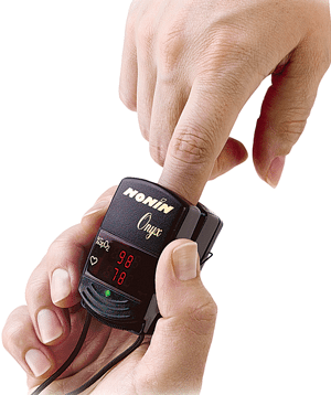
A new non-invasive diagnostic technology could give the single-most important sign of brain health: oxygen saturation. A standard pulse oximeter is clipped onto a finger or earlobe of the subject to measure oxygen levels under the skin. It works by transmitting a beam of light through blood vessels in order to measure the absorption of light by oxygenated and deoxygenated haemoglobin. The information allows physicians to know immediately if oxygen levels in the subject’s blood are rising or falling.
The new device uses a technique called ‘ultrasonic light tagging’ to isolate and monitor an area of tissue the size of a sugar cube located between 1 and 2.5 centimetres under the skin. The probe, which rests on the scalp, contains three laser light sources of different wavelengths, a light detector and an ultrasonic emitter. The laser light diffuses through the skull and illuminates the tissue underneath it. The ultrasonic emitter sends highly directional pulses into the tissue. The pulses change the optical properties of the tissue in such a way that they modulate the laser light traveling through the tissue. In effect, the ultrasonic pulses ‘tag’ a specific portion of the tissue to be observed by the detector. Since the speed of the ultrasonic pulses is known, a specific depth can be selected for monitoring. The modulated laser light is picked up by the detector and used to calculate the tissue’s colour. Since colour is directly related to blood oxygen saturation, it can be used to deduce the tissue’s oxygen saturation.
The measurement is absolute rather than relative, because colour is an indicator of the spectral absorption of haemoglobin and is unaffected by the scalp. Deeper areas could be illuminated with stronger laser beams, but light intensity has to be kept at levels that will not injure the skin.
Given the technology’s current practical depth of 2.5 cm, it is best suited for monitoring the upper layers of the brain. While the technology is designed to monitor a specific area, it could also be used to monitor an entire hemisphere of the brain. Measuring any area within the brain could yield better information about whole-brain oxygen saturation than a pulse oximeter elsewhere on the body.
The author is from Department of Physics, S.L.I.E.T. (Deemed to be University), Longowal, District Sangrur (Punjab)